Literature/202311240735 compressive raman for chemical reactions
- Source: [[@pandya2022Competing oxygen evolution reaction mechanisms revealed by high-speed compressive Raman imaging]]
- Tags: #raman #compressive-raman #oxygen-evolution-reaction
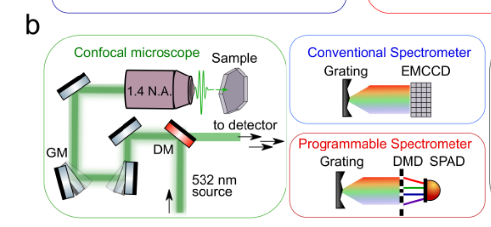
This paper uses Compressive Raman as a tool to get species information on an image. Compared to what is done in [[@gentner2023Compressive Raman microspectroscopy parallelized by single-photon avalanche diode arrays]] (see: 202311240722 Compressive Raman with SPAD Arrays), in this paper there is only a single SPAD used as a detector.
Since they have a point detector, they use a "standard" confocal microscope coupled to a conventional 2D spectrometer or to a compressive Raman system with a DMD and a SPAD.
Interestingly, the paper discusses changes in spectral lines:
We note that on potassium intercalation the computed in-plane 550 cm-1 mode shows relatively little change in spectral position whereas at higher frequencies around 640 cm-1 the computed shifts are larger (~50 cm-1 blueshift)
With compressive Raman, the only way to detect these changes is to perform very narrow scans to crank up the spectral resolution, or to know before hand and explicitly probe two sides of each peak. ($50cm^{-1}$ is around $1nm$ at these wavelengths).
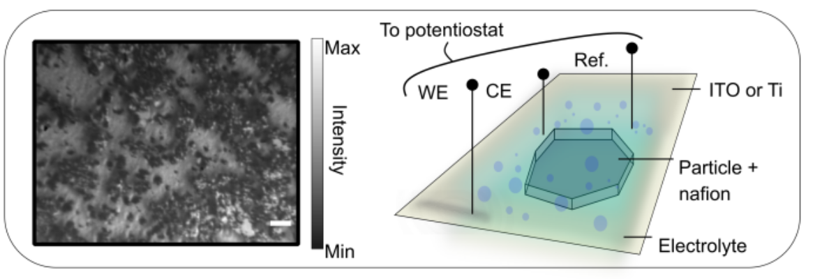
The method yields this type of images of particles undergoing potential cycling:
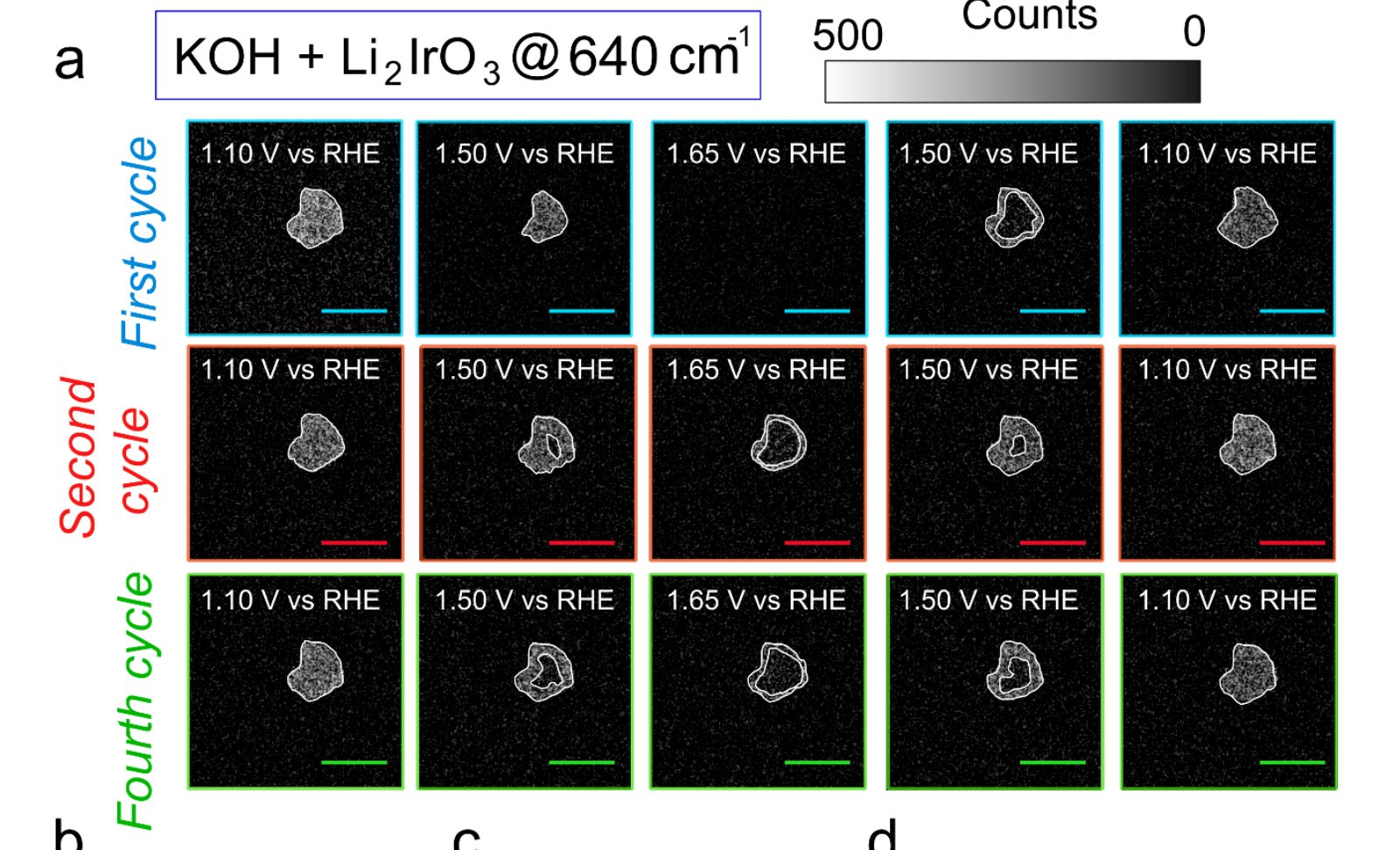
In this case, the authors have selected a specific spectral shift for imaging.
Backlinks
These are the other notes that link to this one.
Comment