202404281926
- Source: [ @vieillard2007 Application of microfluidic chip with integrated optics for electrophoretic separations of proteins ]
- Tags: #Free-flow-electrophoresis #ffe #separation
In this paper they built some microfluidic channel on glass (etching) and covered them with PDMS. I am not sure why they preferred to have glass/PDMS instead of glass/glass (perhaps they didn't have a way of bonding both?)
The glass was soda-lime, the channels had a cross-section of $20\mu m \times 80\mu m$ while the PDMS had a thickness of $1mm$ .
What I found interesting about the paper is that they used the transparent glass to couple a microscope to do real-time monitoring of the signal. They were using fluorescence emission (on an Olympus microscope), but nothing would have prevented them from using scattering or UV-Vis (except the PDMS on top.)
Interestingly, they perform capillary gel electrophoresis and capillary zone electrophoresis measurements, but not free flow electrophoresis . I wonder why they didn't attempt it.
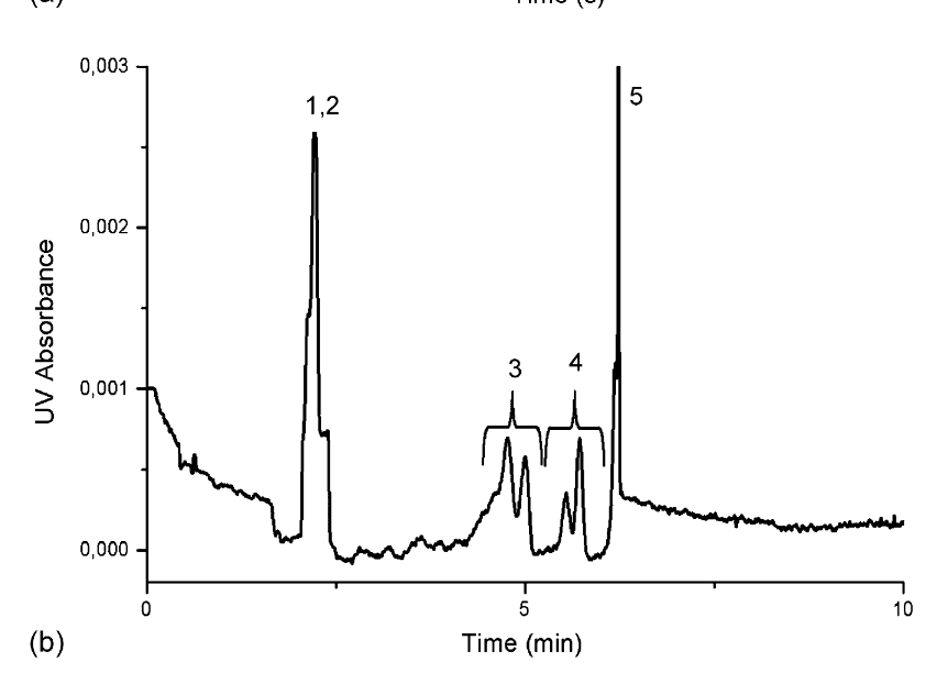
In their system, measurements (and separation) are done as a function of time (see: Time-based separation methods ). On the other hand, it allows for a very simple detection scheme, since a point detector will do.
The schematics of their chips comes from a different paper: [ @mazurczyk2006 A novel concept of the integrated fluorescence detection system and its application in a lab-on-a-chip microdevice ]
Backlinks
These are the other notes that link to this one.
Comment