Sp-iris
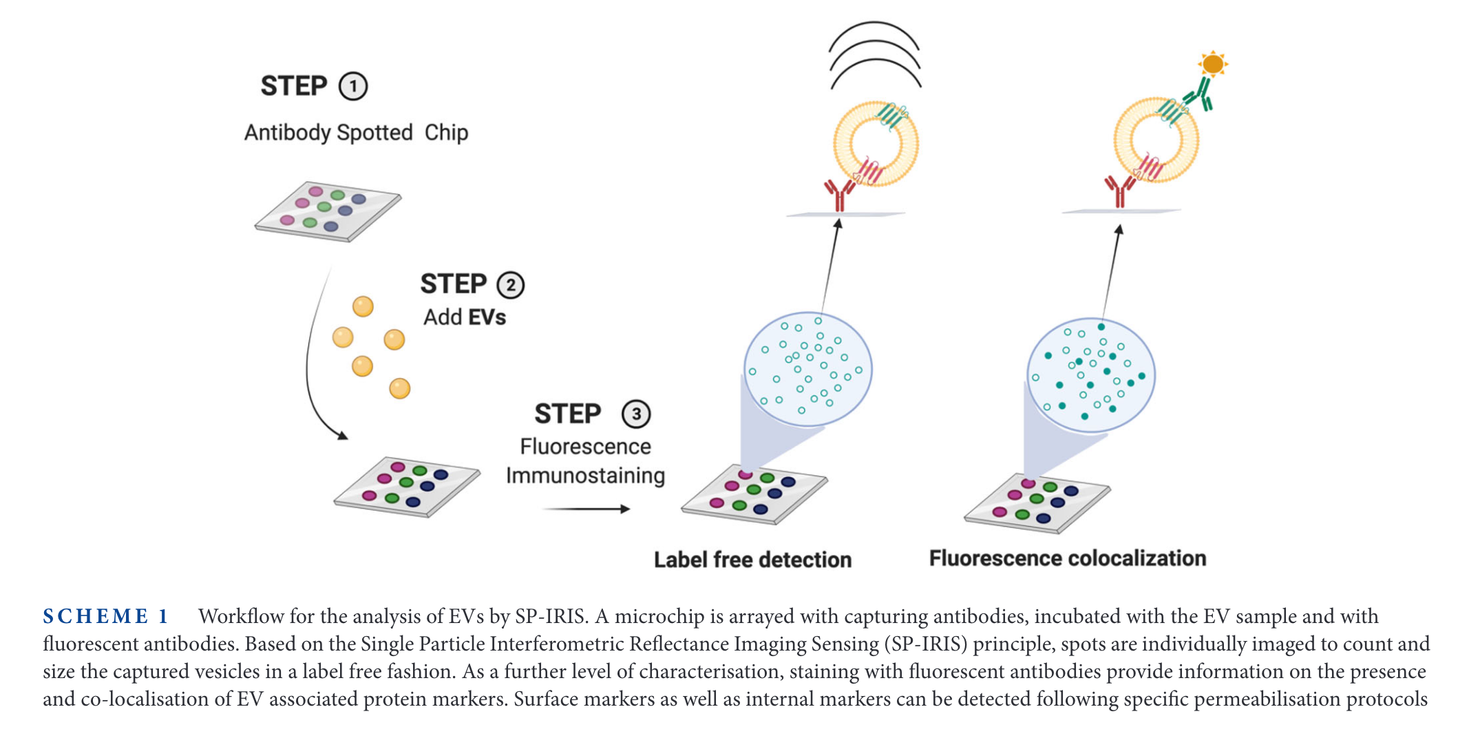
 Scheme of the SP-IRIS setup from [@frigerio2022Comparing digital detection platforms in high sensitivity immune-phenotyping of extracellular vesicles] (and this note).
Single-particle interferometric reflectance imaging sensing is a technique commercialized by NanoView Biosciences under the name ExoView.
They use a microarray functionalized with specific antibodies, which trap particles. Using an interferometric detection system, they can measure the presence (and the size) of the particles at each spot. In a second labelling step (like in Elisa test), the fluorescence signal can be measured.
It is a form of digital detection, with the added size-ability which is purely analog in nature. Since it is an interferometric method, it can work at smaller sizes (extracellular vesicles down to
The interferometric detection seems similar to iSCAT, but I haven't checked the details of the interferometric approach.
A similar measurement approach is that of Simoa, just that in that case, the analytes are trapped by beads and not just by the surface.
Another company producing a relatively similar product is Myriade, under the name Video Drop.
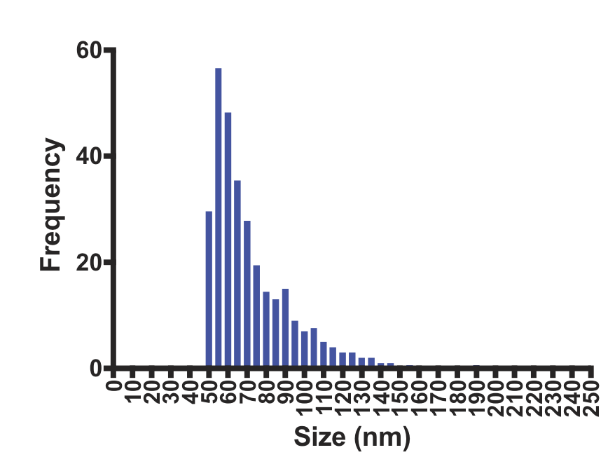
The image below (from [@gori2020Membrane-binding peptides for extracellular vesicles on-chip analysis] and literature/202301141616 Membrane-binding peptides to detect small EVs) can give an idea of the number of particles analyzed in a single-measurement (not sure if it is representative, though)

Integrating the numbers (by eye) gives a total of around 300 particles in the histogram.
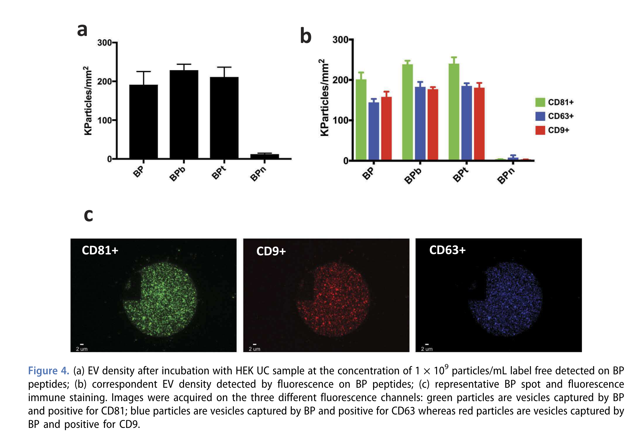
From the same paper, this figure gives a more comprehensive overview:

The image disk is
Backlinks
These are the other notes that link to this one.
